- Cut, Copy, and Paste.
- Selected copy by free hand AOL controlled by four arrow keys available on keyboard or mouse with zoom preview.
- Crop, duplicate, restore
- Resize
- Compression
- Conversion to other format BMP, JPG, TIF, PNG, GIF & PSD
- Flood fill or spray with selected color at selected portion.
- Grid creation; 5X5, 10X10 & 100X100 lines.
- Drawing tool curve, line, square, and circle with node control and provision to change color & thickness of the line.
- Write text in any color or font.
- Pointer to place on an object in four directions with provision to change its color & thickness.
- Eraser works only on line, arrow or on any drawing tool.(not on original image)
- Camera Lucida
- Montage feature to merge stored image together. Useful to Merge different focuses of same image.
- Image stitching.
- Highlighter.
- Pixel by Pixel Correction by key board.
- Multiple image folder with Search facility.
- Filter application on selected area.
|
VIEW
- Zoom in/out
- Zoomed preview
- Rotation at 90, 180,270 or custom
- Image flipping; horizontal or vertical axis
- Intensity histogram.
- Image Information
- Redo/Undo on all operations.
- Ruler in Various units.
- Slide show.
IMAGE PROCESSING
- Background subtraction and contrast enhancement of color or monochrome images
- Arithmatic image functions (Boolean Math; Add, AND, OR, XOR, DIFF, MIN, MAX, +, -, /, *, And Simple).
ROUTINE FILTERS
- Invert, Brightness, Contrast, Hue, Saturation, Blur, Noise Remove, Emboss, Engrave, Gamma R, Gamma G, Gamma B, Yellow, Magenta, Cyan, Mosaic, Smooth, Desaturation, Pseudo Color, Colorize, Oilify, Despeckle, Postarize.
SPECIAL FILTERS & KARNELS
- High Boost, High Spatial, Low Pass Spatial, Ranking (Max, Med, Min), Point detection, Line detection, Homogeneity.
|
EDGE DETECTION
- Laplacing, Sobel, Kirsch, Prewitt Gradient, Shift & Difference, Combine, Contrast Base, Quick, Range And Variance.
MORPHOMETERY
- Skelotizing, Pruning, SKIZ, Histogram Equalization, Histogram Smoothing, Histogram Peak, Histogram Valley, Segmentation by Over/Under and Quantized, Contouring, Dilation / Erosion on Binary, Gray & colored Images, Opening/ Closing on Gray & Binary Images, Special Opening/Closing, Split/Combine Of RGB, YUV, YIQ, XYZ, & HSL, Changing any Image to 1, 4, 8 & 24 Bits, Medial Axis. Transformation, Halftone.
- Image Addition, Image Average, Image Subtraction, Image Multiplication.
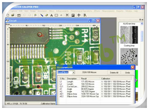
MEASUREMENT
- Spartial calibration
- Line measurements for Distance, Length, Width, Perimeter, Angle, Three Point Radius.
- Area by enclosed line controlled by four arrow keys available on keyboard arrows with zoomed preview.
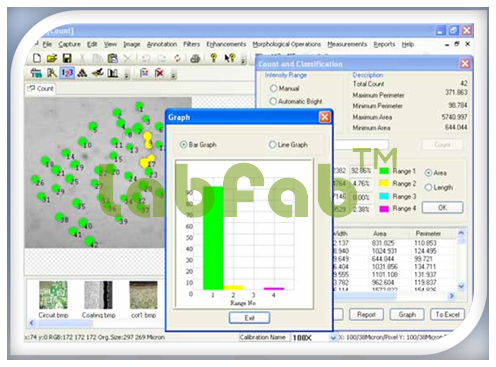
COUNT AND CLASSIFICATION
- Identification of objects in an image, count them, obtain several features measurements. Objects identification by user or automatically. User defined classification on basis of size or intensity.
THRESHOLD PARTICLE MEASUREMENT
- Manual, Auto bright and Auto dark methods to identity intensity range defined object to be measured. Various calculation & measurements available for selected Particle are; Dimensions, Area, Perimeter, Ferrite Length, Min/Max Radius, Thread Length, Thread Width, Fiber Length, Fiber Width.
MORPHOMETERY
- Roundness, Shape, Orientation, Elongation, Equal Circular Diameter, Equal Sphere Volume.
LOCATIONAL
- Centroid X, Centroid Y, Major X1, Major Y1, Minor X1, Minor Y1, Major X2, Major Y, Major X2, Minor Y2, Box X1, Box X2, Box Y, Box Y2 & Box Area.
SEGMENTATION
- Measure area fraction & volume fraction. Identify multiple phases within Microstructure. Also delineate phases from the histogram.
REPORT
- Three options: Direct printout with original image processed Image & Tabular results
- Export to MS Office or Excel for further modification.
|